Smoking Aggravates Keratoconus Severity: A Comprehensive Review
Introduction
Keratoconus (KC) is a progressive corneal disorder characterized by thinning and conical protrusion of the cornea, leading to visual distortion and impaired vision. While genetic predisposition and eye rubbing are well-established risk factors, emerging evidence suggests that environmental factors, including smoking, may exacerbate disease progression. This article explores the relationship between smoking and keratoconus severity, examining biochemical mechanisms, clinical observations, and potential interventions.
Understanding Keratoconus
Keratoconus typically manifests during adolescence and progresses into early adulthood. The condition results from structural weakening of the corneal stroma due to enzymatic degradation of collagen and oxidative stress. Common symptoms include:
- Blurred or distorted vision
- Increased light sensitivity
- Frequent changes in eyeglass prescriptions
- Corneal scarring in advanced cases
Diagnosis relies on corneal topography, pachymetry, and slit-lamp examinations. Treatment options range from rigid gas-permeable contact lenses to corneal cross-linking (CXL) and, in severe cases, corneal transplantation.
The Role of Smoking in Keratoconus Progression
1. Oxidative Stress and Corneal Degradation
Smoking introduces a high concentration of reactive oxygen species (ROS) into the bloodstream, overwhelming the body’s antioxidant defenses. The cornea, being highly vascularized in its limbal region, is susceptible to oxidative damage. Studies indicate that smokers exhibit:
- Increased matrix metalloproteinase (MMP) activity – MMPs degrade collagen, accelerating corneal thinning.
- Reduced antioxidant levels – Glutathione and superoxide dismutase (SOD) are depleted, impairing corneal repair.
- Elevated inflammatory markers – Smoking upregulates pro-inflammatory cytokines (e.g., IL-6, TNF-α), worsening stromal weakening.
2. Impaired Corneal Healing
Nicotine and carbon monoxide in cigarette smoke reduce oxygen supply to ocular tissues, delaying wound healing. This is particularly detrimental for KC patients undergoing corneal cross-linking (CXL), as the procedure relies on riboflavin-induced collagen stabilization under UV light. Smokers may experience:
- Slower epithelial regeneration post-CXL
- Higher risk of complications (e.g., infections, haze formation)
- Reduced treatment efficacy due to persistent oxidative damage
3. Clinical Evidence Linking Smoking to KC Severity
Several studies support the association between smoking and keratoconus progression:
- A 2020 cohort study found that smokers with KC had steeper corneal curvatures and faster progression rates than non-smokers.
- A 2021 meta-analysis reported that smokers were 1.8 times more likely to require corneal transplantation than non-smokers.
- In vivo confocal microscopy revealed greater keratocyte apoptosis in smokers, indicating accelerated stromal degeneration.
Mechanisms Behind Smoking-Induced KC Aggravation
1. Nicotine’s Vasoconstrictive Effects
Nicotine constricts blood vessels, reducing nutrient and oxygen delivery to the cornea. Chronic hypoxia:
- Weakens corneal biomechanics
- Promotes keratocyte apoptosis
- Increases susceptibility to mechanical stress (e.g., eye rubbing)
2. Carbon Monoxide and Oxidative Damage
Carbon monoxide (CO) binds to hemoglobin more efficiently than oxygen, leading to tissue hypoxia. CO also:
- Disrupts mitochondrial function, impairing cellular energy production.
- Enhances ROS generation, exacerbating corneal oxidative stress.
3. Systemic Inflammation and Autoimmune Responses
Smoking triggers systemic inflammation, which may exacerbate KC through:
- Autoantibody production against corneal proteins.
- T-cell-mediated stromal degradation.
Management Strategies for Smokers with Keratoconus
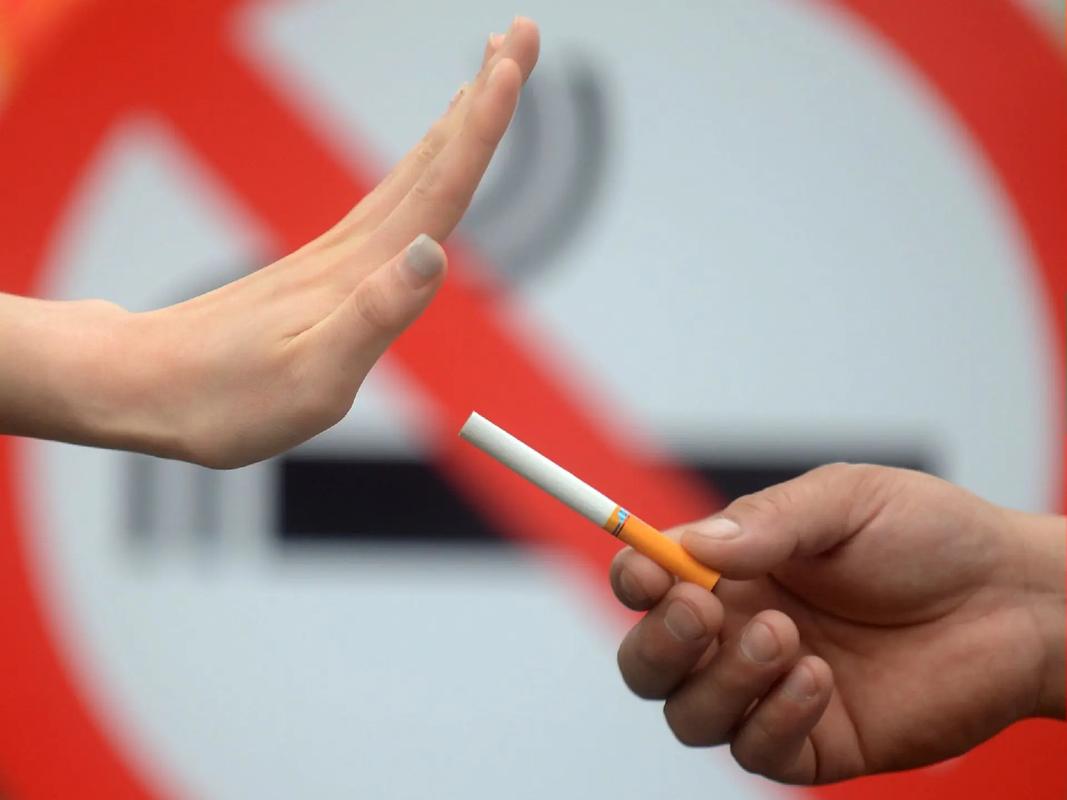
1. Smoking Cessation Programs
The most effective intervention is quitting smoking. Benefits include:
- Reduced oxidative stress within 6–12 months.
- Improved corneal healing post-CXL.
- Slower KC progression compared to active smokers.
2. Antioxidant Supplementation
Patients who smoke may benefit from:
- Vitamin C and E – Neutralize free radicals.
- Omega-3 fatty acids – Reduce inflammation.
- N-acetylcysteine (NAC) – Boosts glutathione levels.
3. Enhanced Monitoring and Early Intervention
Smokers with KC should undergo:
- More frequent corneal topography (every 3–6 months).
- Early CXL to halt progression before severe thinning occurs.
Conclusion
Smoking significantly exacerbates keratoconus severity by increasing oxidative stress, impairing corneal healing, and accelerating stromal thinning. Clinical evidence strongly supports smoking cessation as a critical component of KC management. Ophthalmologists should actively screen for smoking habits in KC patients and provide targeted interventions to mitigate disease progression.
Key Takeaways
- Smoking increases MMP activity and ROS, worsening KC.
- Smokers exhibit faster progression and higher surgical intervention rates.
- Quitting smoking and antioxidant therapy can slow KC advancement.
By addressing modifiable risk factors like smoking, clinicians can improve long-term outcomes for keratoconus patients.
Tags: #Keratoconus #Smoking #Ophthalmology #CornealDisease #OxidativeStress #EyeHealth #MedicalResearch