Title: The Accelerating Peril: How Smoking Fuels the Progression of Honeycombing in Idiopathic Pulmonary Fibrosis
Introduction
Idiopathic Pulmonary Fibrosis (IPF) is a devastating, chronic, and ultimately fatal lung disease characterized by the relentless and irreversible scarring (fibrosis) of the pulmonary interstitium. This process leads to a progressive decline in lung function, causing debilitating shortness of breath and a profound reduction in quality of life. A critical hallmark of advanced IPF, often observed on high-resolution computed tomography (HRCT) scans, is the development of honeycombing. This radiological and pathological pattern describes clustered, cystic air spaces with thickened fibrous walls, typically located in the subpleural regions of the lungs, representing the end-stage architectural destruction of the lung parenchyma. While the "idiopathic" in IPF signifies an unknown cause, numerous environmental and genetic factors are known to influence its initiation and progression. Among these, cigarette smoking stands out as the most significant and modifiable risk factor. A growing body of evidence suggests that smoking does not merely increase the risk of developing IPF; it acts as a powerful accelerator, driving a more rapid progression towards honeycombing and respiratory failure.
The Pathobiology of IPF and Honeycombing
To understand smoking's role, one must first appreciate the complex pathobiology of IPF. The prevailing hypothesis centers on recurrent, microscopic alveolar epithelial cell (AEC) injury in a genetically predisposed individual. In a healthy lung, injury triggers a controlled wound-healing response: the epithelium repairs itself, and any temporary collagen deposited by fibroblasts is eventually remodeled and resolved.
In IPF, this process goes catastrophically awry. The injured AECs, particularly type II cells, undergo aberrant activation and release a plethora of pro-fibrotic mediators, including transforming growth factor-beta (TGF-β), platelet-derived growth factor (PDGF), and connective tissue growth factor (CTGF). These signals recruit fibroblasts and prompt their differentiation into myofibroblasts, the key effector cells of fibrosis. Myofibroblasts proliferate excessively and deposit massive amounts of extracellular matrix (ECM) proteins like collagen, creating stiff, non-functional scar tissue that disrupts gas exchange and lung mechanics.
Honeycombing represents the final, destructive consequence of this unchecked process. It is believed to result from the progressive fibrosis and contraction of the lung parenchyma, which leads to the dilation of terminal and respiratory bronchioles. These dilated airways, filled with mucus and inflammatory cells, and encased in thick fibrous walls, form the characteristic cystic clusters seen on histology and HRCT. The presence and extent of honeycombing are strongly correlated with disease severity, accelerated decline in forced vital capacity (FVC), and increased mortality.
Cigarette Smoke: A Cocktail of Lung Insults
Cigarette smoke is not a single toxin but a complex mixture of over 7,000 chemicals, including reactive oxygen species (ROS), reactive carbonyls, and various carcinogens. This noxious cocktail delivers a multi-pronged assault on the lung that directly exacerbates every stage of IPF pathogenesis.
-
Epithelial Injury and Dysfunction: The primary interface for smoke is the alveolar epithelium. Inhalation causes direct cytotoxicity, damaging and killing AECs. This persistent injury perpetuates the initial trigger of IPF. Furthermore, smoke impairs the innate repair mechanisms of the epithelium, preventing proper healing and creating a state of chronic cellular stress.
-
Oxidative Stress: Cigarette smoke is a tremendous source of exogenous oxidants. It also depletes endogenous antioxidant defenses like glutathione. This creates a state of severe oxidative stress, which activates redox-sensitive transcription factors (e.g., NF-κB) that drive the expression of pro-inflammatory and pro-fibrotic genes. Oxidative stress directly promotes the apoptosis of AECs and stimulates fibroblast proliferation and collagen production.
-
Aberrant Immune Activation and Inflammation: While IPF is not primarily a classic inflammatory disease, innate immune activation plays a crucial role. Cigarette smoke activates alveolar macrophages, shifting them towards a pro-fibrotic (M2) phenotype. These activated macrophages become a potent source of TGF-β and other fibrogenic cytokines. Smoke also disrupts the balance of other immune cells and can trigger the release of damage-associated molecular patterns (DAMPs), further fueling a low-grade but persistent inflammatory response that supports fibrosis.
-
Disruption of Protease/Antiprotease Balance: Smoke increases the release of potent proteolytic enzymes (e.g., neutrophil elastase) from inflammatory cells. These enzymes can directly damage the lung's elastic architecture and activate latent TGF-β, a master switch for fibrosis. Simultaneously, smoke can inactivate protective antiproteases, leading to uncontrolled tissue breakdown and remodeling.
Synergistic Acceleration: How Smoking Fuels Honeycombing
The mechanisms described above create a perfect storm within the already vulnerable IPF lung. Smoking adds jet fuel to the fire of fibrosis, directly accelerating the path toward honeycombing.
- Accelerated Fibroblast Activity: The heightened oxidative stress and cytokine milieu (supercharged by smoke) provide a continuous and intensified signal for fibroblast activation and persistence. This leads to a more rapid and aggressive deposition of ECM, hastening the parenchymal scarring and contraction that culminates in honeycomb cysts.
- Enhanced Tissue Stiffness: The increased ECM deposition directly increases mechanical stress and tissue stiffness. Stiffness itself is a potent pro-fibrotic signal, promoting further fibroblast-to-myofibroblast differentiation in a vicious cycle. This cycle drives the rapid contraction and distortion of lung architecture necessary for honeycomb formation.
- Mucous Plugging and Infection: Cigarette smoke impairs ciliary clearance and increases mucus production. Within the developing microcysts of early honeycombing, this leads to mucus accumulation and plugging. This creates a localized environment prone to bacterial colonization and low-grade infection, which can trigger further epithelial injury, inflammation, and fibrotic progression in a positive feedback loop.
- Earlier and More Extensive Disease: Clinical studies consistently show that IPF patients with a history of smoking present with more severe disease at diagnosis. They exhibit a greater extent of fibrosis and honeycombing on HRCT and experience a faster rate of FVC decline compared to never-smokers. The disease trajectory is simply more aggressive.
Clinical Implications and the Imperative of Cessation
The evidence is unequivocal: continued smoking after an IPF diagnosis is profoundly detrimental. The acceleration of honeycombing progression translates directly to a steeper decline in lung function, earlier onset of respiratory failure, reduced exercise tolerance, and worse survival outcomes.
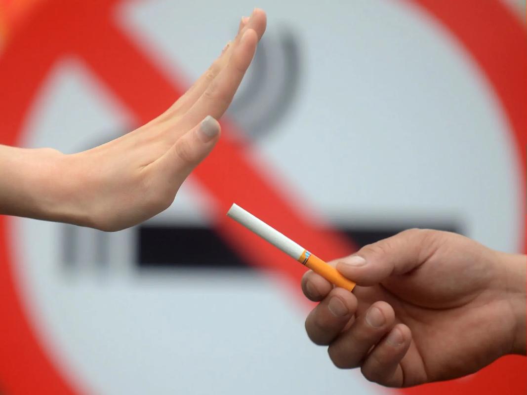
Therefore, smoking cessation is not merely a public health recommendation; it is a critical, non-negotiable component of IPF management. Pulmonologists must prioritize robust smoking cessation counseling, offering behavioral support and pharmacological aids (e.g., varenicline, bupropion, nicotine replacement therapy) to all patients who currently smoke. While quitting cannot reverse established honeycombing, it can potentially slow the relentless drive toward further fibrosis. By removing the primary accelerator, cessation may help decelerate disease progression, helping to preserve remaining lung function for longer and improve the efficacy of antifibrotic medications like pirfenidone and nintedanib.
Conclusion
Idiopathic Pulmonary Fibrosis is a journey along a path of progressive lung scarring, with honeycombing as its devastating destination. Cigarette smoking is the factor that forcefully pushes patients down this path at a terrifying speed. Through direct epithelial injury, profound oxidative stress, and the amplification of core fibrotic pathways, smoke acts as a potent disease accelerator. Understanding this sinister synergy underscores the paramount importance of smoking prevention and cessation. In the fight against IPF, eliminating exposure to cigarette smoke remains one of the most powerful therapeutic interventions available.
Tags: #IdiopathicPulmonaryFibrosis #IPF #Honeycombing #PulmonaryFibrosis #Smoking #CigaretteSmoke #LungDisease #RespiratoryHealth #FibrosisProgression #SmokingCessation #HRCT #COPD #LungDamage #OxidativeStress #TGFbeta