Title: The Inhaled Risk: How Smoking Exacerbates Treatment Failure in Barotrauma-Induced Pneumothorax
Introduction
Pneumothorax, the presence of air in the pleural space causing lung collapse, is a serious medical condition with multiple etiologies. Barotrauma, a form of lung injury induced by significant pressure differentials between the alveolar interior and the surrounding environment, is a well-documented cause. This can occur in scenarios such as mechanical ventilation, scuba diving, or exposure to explosive blasts. The primary and most urgent treatment for a significant pneumothorax is chest tube drainage (thoracostomy), aimed at evacuating air and re-expanding the lung. While this procedure is often successful, a subset of patients experiences treatment failure, characterized by persistent air leaks, incomplete lung re-expansion, or early recurrence. A growing body of clinical evidence points to a critical, modifiable risk factor that significantly increases the likelihood of these complications: tobacco smoking. This article delves into the pathophysiological mechanisms through which smoking compromises pulmonary integrity and healing, directly leading to higher rates of treatment failure in barotrauma pneumothorax.
Understanding Barotrauma Pneumothorax
Barotrauma-related pneumothorax differs from a simple spontaneous pneumothorax in its mechanism of injury. It is not primarily caused by the rupture of a pre-existing bulla (an air-filled sac), but rather by alveolar overdistension and rupture due to high transpulmonary pressures. In mechanical ventilation, this is often a consequence of high tidal volumes or high peak pressures. In diving, it results from breath-holding during ascent, where expanding air trapped in the lungs has no escape route. The initial tear in the alveolar wall creates a bronchopleural fistula (BPF)—an abnormal connection between the bronchial tree and the pleural space. The success of chest tube treatment hinges on the body's ability to swiftly seal this fistula and allow the visceral and parietal pleura to adhere, facilitating lung re-expansion. It is precisely this healing process that smoking profoundly disrupts.
The Multifaceted Assault of Tobacco Smoke on the Lungs

Cigarette smoke is a complex cocktail of over 7,000 chemicals, hundreds of which are toxic and at least 70 known to be carcinogenic. Its impact on the respiratory system is systemic and destructive, creating a lung environment utterly hostile to efficient healing.
-
Impaired Ciliary Clearance and Mucus Hypersecretion: The respiratory epithelium is lined with cilia—microscopic hair-like structures that rhythmically beat to move mucus and trapped particles upward and out of the airways. Tar and other components in tobacco smoke paralyze and destroy these cilia. Concurrently, smoke stimulates goblet cells to produce excessive, thick mucus. This combination leads to mucus plugging, which can obstruct smaller airways. In the context of a pneumothorax, these plugs can prevent distal alveoli from re-inflating even if the major leak is sealed, contributing to atelectasis and treatment failure.
-
Elastase/Antielastase Imbalance and Emphysematous Changes: A pivotal mechanism is the disruption of the protease-antiprotease balance. Smoke inhalation activates pulmonary neutrophils and macrophages, triggering the release of proteolytic enzymes, most notably neutrophil elastase. This enzyme breaks down elastin, a critical protein that provides the lung parenchyma with its elastic recoil. Normally, this activity is checked by alpha-1-antitrypsin, a protective antiprotease. However, components of tobacco smoke oxidize and inactivate this inhibitor. The result is unchecked digestion of the alveolar walls, leading to the destruction of lung architecture and the formation of emphysematous bullae. These areas of damaged, air-filled tissue are structurally weak and highly prone to rupture, both initiating the pneumothorax and creating new or larger fistulas that are difficult to heal.
-
Systemic Inflammation and Oxidative Stress: Smoking induces a state of chronic systemic inflammation. It elevates levels of pro-inflammatory cytokines like TNF-alpha, IL-1, and IL-8, which perpetuate a cycle of tissue injury and edema. Furthermore, the high burden of free radicals in smoke creates significant oxidative stress, damaging cell membranes, proteins, and DNA. This inflamed, oxidative environment impedes every stage of the normal wound healing process—hemostasis, inflammation, proliferation, and remodeling. Fibroblast function, collagen deposition, and angiogenesis (the formation of new blood vessels crucial for repair) are all suppressed.
-
Impaired Immune Defenses: Smoking compromises both innate and adaptive immunity. It disrupts the function of alveolar macrophages, the lungs' first line of defense, and impairs neutrophil chemotaxis (their ability to migrate to the site of injury effectively). This not only increases the risk of secondary infections at the chest tube site or in the pleural space (empyema) but also dysregulates the clean-up of cellular debris necessary for the proliferation phase of healing.
Linking Smoking Directly to Treatment Failure
The pathophysiological changes described above translate directly into clinical treatment challenges:
-
Persistent Air Leak (PAL): This is the most direct manifestation of treatment failure. The combination of a large, friable fistula in emphysematous lung tissue and impaired cellular repair mechanisms means the body cannot effectively seal the bronchopleural fistula. The air leak continues for days or even weeks despite properly placed chest tubes. Studies have consistently identified current smoking as one of the strongest independent predictors of prolonged air leak following lung surgery or traumatic pneumothorax.
-
Incomplete Lung Re-expansion: Even if the major air leak stops, the underlying lung may fail to fully re-inflate. Mucus plugging in the obstructed, cilia-impaired airways traps air distally and prevents alveolar recruitment. The loss of elastic recoil in emphysematous lungs also means the lung lacks the intrinsic "spring" to pull itself back open against the chest wall.
-
High Recurrence Rate: After the chest tube is removed, the lung remains vulnerable. The widespread parenchymal weakness means that even minor pressure changes can cause a new alveolar rupture at a site adjacent to the original or elsewhere in the lung. The failed healing of the initial injury site also leaves a fragile area susceptible to re-rupture.
Clinical Implications and Conclusion
The evidence is clear: active smoking is a powerful driver of poor outcomes in barotrauma pneumothorax. This understanding must inform clinical practice. The management of these patients cannot be limited to the mechanical act of inserting a chest tube. It must include:
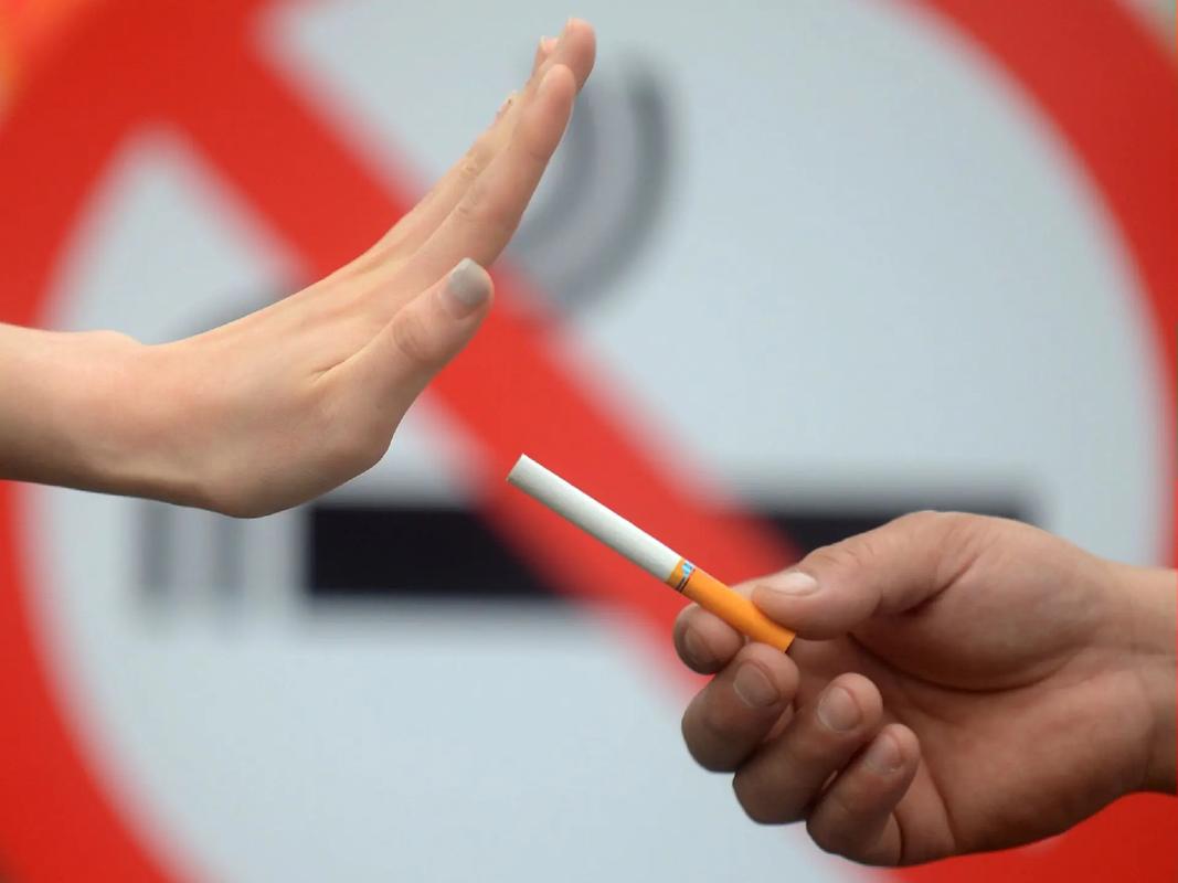
- Aggressive Smoking Cessation Counseling: A diagnosis of pneumothorax represents a "teachable moment." Healthcare providers must urgently and unequivocally advise patients to quit smoking immediately. This is not merely general health advice; it is a critical adjuvant therapy to improve the odds of treatment success.
- Pharmacological Support: The use of nicotine replacement therapy (NRT), varenicline, or bupropion should be considered to manage withdrawal symptoms and support cessation efforts during this critical recovery period.
- Altered Treatment Expectations: Clinicians should anticipate a more complicated and prolonged clinical course in smokers. They may require longer chest tube duration, higher suction levels, or earlier consideration for advanced interventions if conservative management fails, such as video-assisted thoracoscopic surgery (VATS) for pleurodesis or fistula stapling.
In conclusion, the link between smoking and increased failure rates in barotrauma pneumothorax treatment is rooted in the profound damage tobacco inflicts on lung biology. By destroying architecture, inciting inflammation, and crippling repair mechanisms, smoking transforms a manageable injury into a protracted and often recurrent problem. Acknowledging this modifiable risk factor is the first step toward improving patient outcomes through integrated care that addresses both the immediate injury and its underlying cause.


