The Unseen Danger: How Tobacco Use Worsens Outcomes in Pulmonary Mucormycosis
Pulmonary mucormycosis is a formidable and often devastating fungal infection. Caused by a group of molds known as mucormycetes, which are commonly found in soil and decaying organic matter, this disease primarily attacks individuals with compromised immune systems. While the medical community is well-aware of risk factors like uncontrolled diabetes, organ transplantation, and cancer, a critical and modifiable factor often lurks in the shadows: tobacco use. A growing body of evidence and clinical observation points to a troubling conclusion: tobacco use significantly increases the risk of treatment failure in patients battling pulmonary mucormycosis. Understanding this connection is not just an academic exercise; it is a vital step towards improving survival rates and patient outcomes.
To grasp how tobacco influences this rare infection, we must first understand the unique battlefield that is the human lung. Our respiratory system has a sophisticated multi-layered defense system. Tiny hair-like structures called cilia line our airways, constantly beating in a coordinated wave to move mucus, trapped particles, and pathogens up and out of the lungs. This "mucociliary escalator" is a first line of defense. Alveolar macrophages, specialized immune cells, patrol the deepest air sacs, engulfing and destroying invaders that make it past the initial barriers.
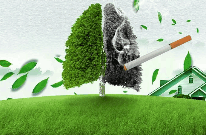
Now, introduce tobacco smoke—a toxic cocktail of thousands of chemicals. Smoking systematically dismantles these defenses. It paralyzes the cilia, halting the essential cleansing motion of the mucociliary escalator. It also damages and kills the cilia, leading to a permanent impairment of this clearance mechanism. Simultaneously, tobacco smoke overwhelms and destroys alveolar macrophages, blunting their ability to recognize and attack fungal spores. In essence, smoking creates a "perfect storm" within the lungs: it provides a direct delivery system for fungal spores deep into the respiratory tract and then disables the very mechanisms designed to eliminate them. This creates a fertile ground for initial infection, a concept known as increased susceptibility to pulmonary mucormycosis.
However, the problem extends far beyond the initial infection. The real crisis unfolds during treatment, where tobacco's insidious effects directly contribute to a higher pulmonary mucormycosis treatment failure rate. The cornerstone of managing this aggressive infection is a two-pronged approach: aggressive surgical debridement to remove dead and infected tissue, and prompt administration of potent antifungal medications, most notably amphotericin B.
This is where tobacco's impact becomes critically evident. Let's break down the key mechanisms:
1. Compromised Blood Flow and Tissue Hypoxia: Mucormycetes are notorious for their ability to invade blood vessels. The fungi grow into the arteries, causing blood clots (thrombosis) that cut off blood supply to surrounding tissue. This leads to tissue death (necrosis), which is a hallmark of the disease. Tobacco use is a primary driver of peripheral vascular disease. The chemicals in smoke damage the endothelial lining of blood vessels, promote inflammation, and cause vasoconstriction (narrowing of blood vessels). In a patient with pulmonary mucormycosis, who already has compromised blood flow due to fungal invasion, pre-existing tobacco-induced vascular damage is a catastrophic combination. It severely limits blood supply to the infected area, creating an environment of profound hypoxia (low oxygen).
This hypoxia serves a dual negative purpose. First, it further promotes tissue necrosis, making the infection more extensive and difficult to surgically eradicate. Second, and perhaps more critically, it impairs the delivery of antifungal drugs. Antifungal agents travel through the bloodstream to reach the site of infection. If the blood vessels are damaged, constricted, and blocked, the medication cannot penetrate the infected tissue in effective concentrations. This directly leads to sub-therapeutic drug levels at the precise location where they are needed most, rendering the pharmacological treatment less effective and directly contributing to a poor response to pulmonary mucormycosis therapy.
2. Immunosuppression Beyond Macrophages: While the damage to alveolar macrophages is a key initial event, tobacco's immunosuppressive effects are systemic and complex. Smoking alters the function of neutrophils, another crucial type of white blood cell essential for fighting fungal infections. It also disrupts the balance of cytokines, the signaling molecules that coordinate the immune response. This results in a dysregulated and inefficient immune attack against the mucormycetes. Even with the best drugs and surgery, a functional immune system is a crucial partner in clearing an infection. In a tobacco user, this partner is weakened and confused, creating a significant obstacle to recovery and increasing the overall burden of fungal infection in the body.
3. The Challenge of Wound Healing: For patients who undergo life-saving surgery, the next hurdle is healing. Surgical resection of a part of the lung is a massive trauma to the body. Successful recovery depends on robust tissue repair and regeneration. Tobacco smoke is profoundly detrimental to wound healing. The carbon monoxide in smoke reduces the oxygen-carrying capacity of blood, while nicotine causes vasoconstriction, both of which deprive healing tissues of essential oxygen and nutrients. Furthermore, smoking interferes with collagen production, the fundamental building block for new tissue. Consequently, post-surgical complications such as delayed healing, bronchopleural fistula (a leak between the lung and the air space around it), and prolonged air leaks are significantly more common in smokers. These complications can delay further treatment, require additional interventions, and provide a continued nidus for infection, all of which elevate the risk of mortality from mucormycosis.
The Clinical Evidence and Patient Trajectory
The correlation is stark in clinical settings. Patients with a history of tobacco use who develop pulmonary mucormycosis often present with more extensive disease. Their clinical course is frequently more aggressive, with a faster progression of symptoms like cough, chest pain, fever, and hemoptysis (coughing up blood). When reviewing cases, physicians often note that the window for effective intervention is narrower for these patients.
The term managing pulmonary mucormycosis in smokers has become synonymous with a more complex and fraught clinical pathway. Treatment plans must account for their compromised physiology. Surgeons may find the tissue planes more difficult to navigate due to smoking-related lung disease like emphysema, and anesthesiologists face heightened risks due to poor lung function. The impact of smoking on antifungal therapy efficacy is a constant concern, often leading to prolonged courses of intravenous medication and the need for therapeutic drug monitoring, where possible.

Perhaps the most critical long-term consideration is the link between tobacco use and recurrent fungal infection. Even if a patient survives the initial catastrophic illness, their underlying lung environment remains altered by tobacco. The damaged architecture and persistent immune dysfunction mean they are at a higher risk for future bacterial and fungal infections, creating a vicious cycle of poor respiratory health.
A Call to Action: Integrating Smoking Cessation into Care
The evidence is clear: addressing tobacco use is not a secondary public health message but an integral component of treating pulmonary mucormycosis. For patients diagnosed with this infection, smoking cessation becomes a non-negotiable, urgent medical intervention. The moment of diagnosis, while frightening, can also be a powerful "teachable moment," providing immense motivation for patients to quit.
Healthcare teams must integrate robust smoking cessation support directly into the treatment protocol. This includes:
- Behavioral Counseling: Providing consistent, empathetic counseling from doctors, nurses, and respiratory therapists.
- Pharmacotherapy: Offering nicotine replacement therapy (gums, patches, lozenges) or medications like bupropion and varenicline to manage withdrawal symptoms, even during hospitalization.
- Long-term Support: Connecting patients with resources for long-term maintenance of a smoke-free life.
Improving the survival rate of pulmonary mucormycosis in immunocompromised patients requires a holistic attack on all risk factors. While we cannot instantly reverse diabetes or undo chemotherapy, smoking is a variable we can and must influence. Every cigarette not smoked is a step towards restoring lung defenses, improving blood flow, and giving life-saving antifungal drugs a fighting chance to reach their target.
In conclusion, the relationship between tobacco and pulmonary mucormycosis is a deadly synergy. Tobacco smoke prepares the lung for invasion, sabotages the body's defenses, undermines surgical and medical treatment, and hinders recovery. It is a major, modifiable driver of the devastatingly high pulmonary mucormycosis treatment failure rate. For any patient facing this diagnosis, the message must be unequivocal: quitting tobacco is as crucial as the first dose of antifungal medicine. It is, without exaggeration, a decision that could mean the difference between life and death.


